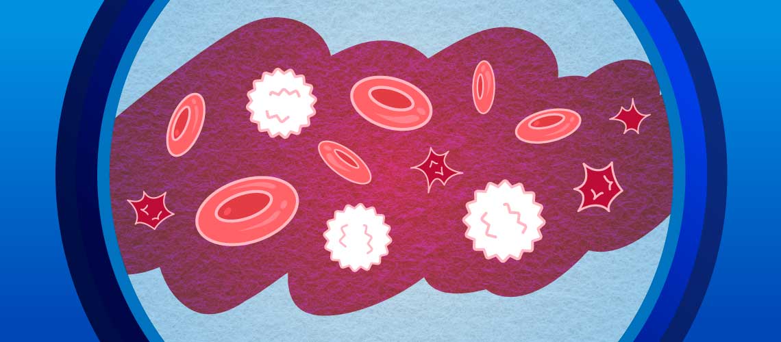
Rethinking the Left Shift: A Data-Driven Approach to Inflammatory Diagnostics
A complete blood count (CBC) remains one of the most fundamental diagnostic tools in small animal practice. However, many clinicians use only half of this test's utility when inflammation is suspected, relying solely on the numerical results. Pairing CBC results with blood smear evaluation is essential, especially when a left shift is present. The combination of quantitative and qualitative data enables veterinarians to differentiate regenerative from degenerative responses, identify toxic neutrophil changes, assess the severity of inflammation, and obtain an earlier understanding of diagnostic direction from critical clues, leading to better patient outcomes. Without examining both the numbers and the morphology, important diagnostic clues may be missed, leading to incomplete or suboptimal case management.
Understanding the Inflammatory Pattern: Regenerative vs. Degenerative Left Shift
The first step in understanding the pattern of inflammation on a CBC is to fully examine the neutrophil's role. Neutrophils are the first responders in acute inflammation, and the timing around their production, maturation, and release into the bloodstream provides a real-time assessment of the bone marrow’s response to disease.
A regenerative left shift occurs when mature neutrophils are accompanied by an increased number of bands, indicating that the bone marrow is keeping pace with demand. This pattern typically reflects an adequate bone marrow response and is typically seen with bacterial infections, sterile inflammatory processes, immune-mediated disease, and early neoplastic inflammation. The presence of more mature, segmented than immature neutrophils such as bands distinguishes a regenerative response and generally carries a more favorable prognosis than degenerative left shift.1,2
In contrast, a degenerative left shift—defined by more immature forms (bands or earlier precursors) than segmented neutrophils—signals that demand exceeds bone marrow capacity. This pattern usually indicates severe inflammation, sepsis, endotoxemia, or overwhelming infection. Degenerative left shifts are also associated with increased mortality risk in critically ill dogs and cats, often due to euthanasia.1,2 Early recognition and distinction are crucial because treatment urgency, monitoring frequency, and prognosis differ dramatically between regenerative and degenerative responses.
Common Causes of Left Shift and Inflammation
While bacterial infection is one of the most common inciting causes for a left shift, it is just one part of a much longer differential list. Disease categories commonly associated with left shift include:
-
Bacterial and fungal infections: pyometra, septic peritonitis, pneumonia, abscesses, discospondylitis, and fungal pneumonias
-
Immune-mediated diseases: immune-mediated hemolytic anemia (IMHA), immune-mediated thrombocytopenia (ITP), and polyarthritis
-
Neoplasia: particularly those producing necrosis or secondary infections
-
Sterile inflammation: pancreatitis, trauma, heatstroke, ischemia, foreign bodies
-
Endotoxemia and systemic inflammatory response syndrome (SIRS): conditions driving marrow exhaustion and degenerative shifts3
Because inflammatory CBC patterns are nonspecific, morphology and clinical correlation are essential for identifying the underlying cause.
Why Morphology Matters
CBC analyzers provide a rapid quantitative overview, but only a blood smear reveals the morphology necessary to accurately interpret inflammatory changes. Key findings to evaluate on smears include:
1. Immature neutrophils
The presence of immature neutrophils, such as bands, metamyelocytes, and myelocytes, confirms the presence and degree of a left shift. Automated analyzers may flag these immature cells, but manual confirmation on a blood smear is often necessary, especially when cell shapes are abnormal.
2. Toxic changes
Toxic neutrophils—characterized by Döhle bodies, cytoplasmic basophilia, vacuolation, and toxic granulation—are often seen before a left shift and are considered sensitive markers of inflammation.4 The presence of marked toxic change usually indicates a more severe and systemic process, often supporting the need for hospitalization, antimicrobial therapy, and aggressive monitoring.
3. Reactive lymphocytes and atypical cells
Blood smear evaluation can distinguish true lymphocytosis from reactive causes (e.g., vaccination, infection) versus neoplastic conditions like lymphoma or leukemia—diagnoses that cannot be made from numerical data alone.
4. Platelet clumping
Finally, blood film evaluation corrects an erroneous diagnosis of thrombocytopenia caused by platelet clumping in automated CBC analyzers, where clumps of two or three platelets may be counted as one or larger clumps may not be counted at all.
Correlating CBC Findings With Clinical Signs and Diagnostics
However, we cannot interpret a left shift in isolation: It's essential to correlate with the patient's clinical condition. A patient with band neutrophilia but normal vital signs may tolerate outpatient monitoring. But, the same CBC pattern in a patient with fever, hypotension, or tachycardia suggests possible sepsis requiring hospitalization and additional diagnostics such as:
-
Blood cultures
-
Imaging to look for the source
-
Coagulation testing
-
+/- lactate measurement
-
+/- acute-phase proteins (e.g., C-reactive protein)
The Role of Automated Analyzers in Real-Time Care
Today's in-clinic hematology analyzers have transformed the speed and precision with which blood counts and inflammatory changes are assessed. Advanced veterinary analyzers can now:
-
Flag left shifts and identify immature granulocytes within minutes.
-
Identify bands with greater accuracy using multi-angle light-scattering or flow cytometry-based technologies.
-
Detect abnormal cell populations that prompt further blood smear review.
-
Integrate with automated microscopy systems for on-screen morphology evaluation.
These innovations reduce the chance of missing subtle hematologic abnormalities and support real-time decisions for critical patients. Emerging artificial intelligence–assisted systems also show promise in improving consistency and accuracy in detecting toxic changes and identifying immature cells.
Toxic and Immature Neutrophils: Implications for Severity and Prognosis
When toxic and immature neutrophils are identified, the degree of left shift and the presence of toxic change can also strongly correlate with disease severity.
-
Mild toxic change may appear with moderate inflammation.
-
Marked toxic change is usually associated with sepsis, systemic inflammation, and poorer outcomes.
-
And pronounced degenerative shifts usually indicate bone marrow exhaustion and increased risk of patient decompensation.
Recognizing these patterns early allows veterinarians to guide client communication, triage appropriately, and pursue aggressive therapy when warranted.
Translating Left Shift Findings into Better Clinical Outcomes
A left shift is much more than an abnormal CBC—it is a flashlight onto the body’s inflammatory response. Only by correlating CBC data with blood film morphology, clinical signs, and advanced analyzer insights can clinicians fully interpret the inflammatory pattern and response. Distinguishing regenerative from degenerative left shifts, identifying toxic changes, and pairing these findings with a patient’s clinical condition enables timely and accurate decision-making. The recognition and interpretation of a left shift ultimately clarify incremental diagnostics, thereby strengthening patient outcomes.
References:
-
Burton, A.G., Harris L.A., Owens, S.D., & Jandrey, K.E. (2013). The prognostic utility of degenerative left shifts in dogs. Journal of Veterinary Internal Medicine, 27(6), 1517-1522. https://doi.org/10.1111/jvim.12208
-
Burton, A.G., Harris, L.A., Owens, S.D., & Jandrey, K.E. (2014). Degenerative left shift as a prognostic tool in cats. Journal of Veterinary Internal Medicine. 28(3), 912-917. https://doi.org/10.1111/jvim.12338.
-
Gori, E., Pierini, A., Lippi, I., Lubas, G., & Marchetti, V. (2021). Leukocytes ratios in feline systemic inflammatory response syndrome and sepsis: A retrospective analysis of 209 cases. Animals, 11(6), 1644. https://doi.org/10.3390/ani11061644
-
Segev, G., Klement, E., & Aroch, I. (2006). Toxic neutrophils in cats: Clinical and clinicopathologic features and disease prevalence and outcome—A retrospective case control study. Journal of Veterinary Internal Medicine, 20(1), 20-31. https://doi.org/10.1892/0891-6640(2006)20[20:TNICCA]2.0.CO;2