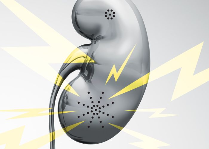

A Veterinary Deep Dive into Proteinuria
Proteinuria in pets refers to the presence of any protein, such as albumin, globulins, mucoproteins, and Bence-Jones proteins, in urine.1 In healthy patients, only trace amounts of albumin are excreted in the urine. However, with some types of kidney dysfunction, they allow the passage of larger protein molecules as part of the urine filtrate. This is a significant clinical finding and requires further investigation and treatment.
What Is Proteinuria?
In veterinary practice, protein in the urine is diagnosed through various measurements, including the urine protein to creatinine ratio (UPC). Dogs are considered nonproteinuric (normal) with a UPC < 0.2, borderline proteinuric at a UPC of 0.2 to 0.5, and significantly proteinuric with a UPC of > 0.5. Cats are also considered nonproteinuric with a UPC < 0.2, borderline proteinuric at a UPC of 0.2 to 0.4, but significantly proteinuric at a UPC of > 0.4.2
Proteinuria Pathophysiology
Reviewing the pathophysiology can steer us to differential diagnoses. With proteinuria, it's helpful to understand the function of glomerular filtration barrier (or glomerular membrane). This membrane has three layers: the fenestrated endothelium, the glomerular basement membrane, and the podocytes. The last layer is most important in preventing proteinuria—its slit diaphragm selects the passage of proteins by both size and charge, and few negatively-charged proteins larger than albumin (69,000 daltons) can pass through it.2 Proteinuria develops when podocytes dysfunction or become overwhelmed with the amount of protein for the kidneys to filter and absorb.
Types of Proteinuria
Proteinuria can be divided into three categories: prerenal, renal, and post-renal.
Prerenal proteinuria is most associated with the overwhelm and overload scenario. Specifically, this can occur when the proximal tubule is overwhelmed and cannot reabsorb low molecular weight proteins like hemoglobin, myoglobin, immunoglobulins, and Bence-Jones proteins.3 This type of proteinuria can often be caused by autoimmune diseases, multiple myeloma, and syndromes that create immune complexes.
Alternatively, renal proteinuria is typically caused by dysfunction in the glomerular filtration barrier, tubular reabsorption like with Fanconi syndrome, or damage to the interstitial tissues. Diseases like glomerulonephritis and amyloidosis can create permanent and persistent damage to the glomerular filtration barrier and cause the highest proteinuria levels in pets.4 Other sources of inflammation, like pyelonephritis, leptospirosis, renal neoplasia, and nephroliths, can cause renal interstitial disease.
Finally, post-renal proteinuria is due to protein in the urine from sources in the urinary tract distal to the kidney, such as urinary tract infections, vaginitis/prostatitis, and hemorrhage. This form of proteinuria can resolve when the underlying cause is identified and treated.
Detecting Proteinuria in Pets
One mistake some veterinarians make is screening for proteinuria solely in patients diagnosed with kidney disease. However, proteinuria in pets can be associated with many different disease states and breeds that are more predisposed to protein-losing disease.5 If routine blood work is indicated or a patient is at risk due to breed or disease, a urinalysis and evaluation of proteinuria should also be performed concurrently.
Persistent protein in the urine is recognized using three serial urinalyses with negative sediments more than two weeks apart. Note that hyaline casts are a consequence of protein in the urine and should not be considered as a part of an active sediment in this scenario.3 Recommended testing includes:
- Dipstick colorimetric method: This is the first screening test for clinicians. When the reagent strip is dipped in urine, a color change will occur if albumin (and other proteins) is present. However, these sticks were created for human urine, which is not usually as concentrated as dog or cat urine; therefore, false positives are possible if the urine is highly concentrated, alkaline, or pigmented.3 Conversely, false negatives are more common in acidic urine and with feline patients. Quantification of urine protein with a urine protein:creatinine ratio provides greater accuracy than dipstick alone.
- Urine protein to creatinine ratio (UPC): This test is recommended for any dog or cat with a trace or greater urine dipstick for protein.10 The UPC ratio is a quantitative measure of all urine proteins. Urine protein and urine creatinine are measured together and the UPC ratio normalizes for variations in urine concentration.
Clinical Significance of Proteinuria
When proteinuria is detected and confirmed, it's also important to look at its severity. If the patient has borderline proteinuria, a 3-month recheck is recommended1. However, if the proteinuria is significant, or the patient is hypoalbuminemic, the veterinary team should move forward with a more complete diagnostic workup. If severe proteinuria is detected, timely diagnostics are indicated.
Proteinuria should never be ignored as it can create significant clinical consequences in the patient. Renal loss of protein can contribute to hypoalbuminemia, coagulation abnormalities, changes in cellular immunity, minerals, electrolyte metabolism, and even hormone status7, and is closely related to prognosis. Multiple studies show that proteinuria is closely related to reduced survival in azotemic and non-azotemic dogs and cats.6-9
Diagnosing Underlying Causes of Proteinuria
The clinical workup encompasses several different components. Begin with a comprehensive medical history, including previous vaccines, travel, and parasite-preventive use. Then ask the pet owner for specific observations around appetite, thirst, energy level, urination amount and frequency, gastrointestinal signs, and other abnormal behaviors. Paired with results from a complete physical exam, focusing on ophthalmic changes, joint swelling, abnormal abdominal palpation, and the presence of fever, you may be able to better understand the cause of the proteinuria.
The minimum database for a proteinuric patient includes a complete blood count (CBC), chemistry panel with electrolytes, blood pressure, and urine culture. Additionally, for dogs, include heartworm testing and tick titers, leptospirosis PCR if the patient demonstrates polyuria and polydipsia, and hyperadrenocorticism if history and physical support the diagnosis. If indicated, feline leukemia virus (FeLV) and feline immunodeficiency virus (FIV) testing should be added for cats, along with pancreatic-specific lipase and additional thyroid testing. Imaging such as thoracic radiographs, abdominal ultrasounds, and CT scans can also help identify structural abnormalities.
Using proteinuria testing as a jumping-off point can allow you to move forward with other testing and diagnostics, and give you a look into the health of a pet. Earlier diagnosis of the underlying cause allows for earlier treatment and the prospect of a better outcome.
References:
- Grauer, G. F. (2011). Proteinuria: Measurement and Interpretation. Topics in Companion Animal Medicine, 26(3), 121-127.
- Littman, M.P., Daminet, S., Grauer, G.F., Lees, G.E. and van Dongen, A.M. (2013), Consensus Recommendations for the Diagnostic Investigation of Dogs with Suspected Glomerular Disease. J Vet Intern Med, 27: S19-S26. https://doi.org/10.1111/jvim.12223
- Harley, L., & Langston, C. (2012). Proteinuria in dogs and cats. The Canadian Veterinary Journal, 53(6), 631-638.
- Lees GE, Brown SA, Elliot J, Grauer GF, et al. Assessment and management of proteinuria in dogs and cats: 2004 ACVIM forum consensus statement (small animal) J Vet Intern Med. 2005;19:377–385.
- Littman MP, Goldstein RE, Labato MA, et al. ACVIM small animal consensus statement on Lyme disease in dogs: Diagnosis, treatment, and prevention. J Vet Intern Med. 2006;20:423–434.
- Whittemore JC, Gill VL, Jensen WA, et al. Evaluation of the association between microalbuminuria and the urine albumin-creatinine ratio and systemic disease in dogs. J Am Vet Med. 2006;229:958–963.
- Bernard DB. Extrarenal complications of the nephrotic syndrome. Kidney Int. 1988;33:1184.
- Cook AK, Cowgill LD. Clinical and pathologic features of protein-losing glomerular disease in the dog: A review of 137 cases (1985–1992) J Am Anim Hosp Assoc. 1996;32:313–22.
- Jacob F, Polzin DJ, Osborne CA, et al. Evaluation of the association between initial proteinuria and morbidity rate or death in dogs with naturally occurring chronic renal failure. J Am Vet Med Assoc. 2005;226:393–400.
- Vaden SL, Elliott J. Management of Proteinuria in Dogs and Cats with Chronic Kidney Disease. Vet Clin N Amer Sm Anim Pract. 2016 Nov;46(6):1115-1130


