

Under the Surface: What Dental Radiographs Reveal in Veterinary Patients

A thorough oral examination is an essential first step in evaluating a dog's dental health. However, it is inherently limited to what can be observed and palpated on the tooth and gingival surface. Visible signs like gingival inflammation, bleeding, or recession can indicate underlying issues, but they provide little information about the condition of the alveolar bone. Even experienced veterinarians may overlook critical changes such as bone loss, abscesses, or osteolysis without radiographs.
A study of 226 dogs found that full-mouth intraoral dental radiographs confirmed clinical findings in all dogs, and radiographs showed that 92% of the dogs had underlying bone loss. Additionally, radiographs of teeth without visible oral lesions found clinically important findings in 27.8% of the cases.1 A similar study in cats found that 42% of the cases had clinically important findings in visibly normal teeth.2
Better dentals begin with a better workflow. Learn more about adding dental to IDEXX Web PACS.
The American Animal Hospital Association (AAHA) guidelines assert that an anesthetized oral examination with intraoral radiographs is necessary for a complete oral health assessment. Forgoing dental radiographs can delay the diagnosis and treatment of alveolar bone disease, allowing painful conditions to progress unnoticed.3
Radiographic Interpretation
Radiographic positioning is extremely important to create consistency between images and patients and increase the accuracy of interpretation. All dental radiographs should be acquired with the patient under general anesthesia, facilitating proper positioning while maintaining patient and staff protection.
A complete intraoral radiograph series includes the following regions:4
- Occlusal view of maxillary canines and incisors
- Right maxillary lateral canine image
- Right maxillary premolars/molars
- Left maxillary lateral canine image
- Left maxillary premolars/molars
- Occlusal view of mandibular incisors and canines
- Right mandibular lateral canine image
- Right mandibular premolars/molars
- Left mandibular lateral canine image
- Left mandibular premolars/molars
Understanding normal radiographic anatomy for the patient's age is paramount before addressing radiographic abnormalities. For example, as patients age, the maturation of the teeth will affect the size of the pulp chamber and root canal. Intraoral dental radiographs allow for visualization of the underlying bone and root structures for a complete evaluation of periodontal disease, endodontic disease, oral trauma, tooth resorption, and neoplasia. All of this information is imperative for treatment, surgical planning, and monitoring patients' dental health over time. 4
Radiographic Assessment of Disease
An oral exam is just the starting point for dental health. Radiography is necessary to rule out underlying issues, including:
Periodontal Disease
Periodontal disease is divided into four stages based on clinical and radiographic findings. Stage 1 has no reversible bone loss, Stage 2 has less than 25% bone loss, Stage 3 has 25-50% bone loss, and Stage 4 has greater than 50% bone loss. Radiographic findings associated with periodontal disease include widening of the periodontal ligament space, loss or indistinct lamina, decreased alveolar bone density, and alveolar bone loss.4
Types of bone loss include:
- Horizontal bone loss: Alveolar bone lysis along adjacent teeth, buccal and lingual plates of bone may be partially resorbed
- Vertical bone loss: Alveolar bone lysis that extends apically from the alveolar margin
- Furcation exposure: Bone loss at the root junction of multi-rooted teeth, identified as radiolucent bone lysis at the furcation
- Alveolar bone expansion: Expansile osteolysis of the buccal alveolar bone that primarily affects feline maxillary or mandibular canine teeth
Determining the type(s) of alveolar bone loss is clinically significant as it will determine if treatment or extraction is indicated.5

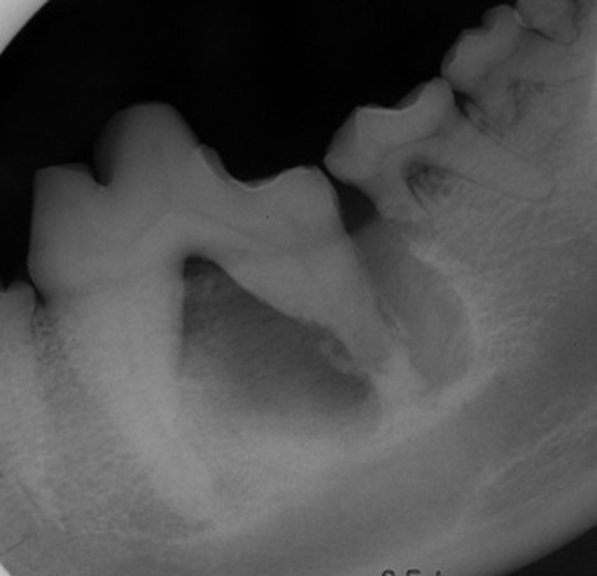
Horizontal bone loss with furcation exposure at the left maxillary fourth premolar (208)
Vertical bone loss with furcation exposure at the distal root of left mandibular fourth premolar (308)
Severe vertical bone loss with furcation exposure and periapical lucency at the distal root of the left mandibular first molar (309) with resultant class II perio/endo lesion
Endodontic Disease
Endodontic disease can be suspected clinically when fractured or discolored teeth or a parulis is present. However, radiographs are essential for determining the extent of the disease. Endodontic disease refers to conditions affecting the root canal, apex or periapex. Radiographic evaluation of the crown, root, lamina dura, dentin, and pulp are crucial. Treatment options depend on radiographic findings and are important for monitoring progression over time.4,5
Radiographic findings include:
- Surrounding tooth changes: Periapical lucency, loss of lamina dura, diffuse alveolar bone sclerosis
- Tooth changes: Wide root canal compared to contralateral tooth, periapical lucency, periapical sclerosis, inflammatory root resorption and internal resorption
Periodontal-Endodontic Disease
Radiographic changes to both the periodontal space and the tooth can happen as an extension of periodontal disease to infection of the root canal (perio-endo lesion), or endodontic disease can progress to periodontal disease (endo-perio lesion). Radiographic findings from both groups will be identified on the same tooth. 4,5
Class II perio/endo lesion at 309. Small well defined periapical lucency at mesial root 309. Large well defined periapical lucency with osteosclerotic rim and inflammatory root resorption distal root 309 with resultant root fracture at the mesial root of 310.
Tooth Resorption
Tooth resorption is seen in both canine and feline patients. In feline patients there are three major types. Type I is a lucency in the tooth that is not replaced by bone dense tissue, whereas type II is a loss of the PDL and a replacement of tooth substance with bone dense tissue. Type III is a combination of both. The American Veterinary Dental College (AVDC) also stages tooth resorption as to its severity and extent of lesion. Stage 1 involves the enamel or cementum, stage 2 involves the dentin, stage 3 involves the pulp, stage 4 destroys significant amount of crown predisposing it to fracture and stage 5 is a lesion that has destroyed the entire crown.6 Tooth resorption in dogs is very common in 53.6% of dogs. There are seven types of tooth resorption in dogs with external replacement resorption being the most common in 34% of dogs and external inflammatory resorption in 25.9%. There is a positive association between tooth resorption and age and larger size of dog. External replacement resorption usually begins at the early premolars and progresses to other teeth.
Tooth resorption/periodontitis in the cat. 307 (stage2, type II), 308-309 (stage 4a, type III) and horizontal bone loss with furcation exposure at 308-309.
Squamous cell carcinoma of left rostral to mid mandible in the cat.
Moth-eaten osteolysis of left mandible from midline to what can be seen of mid mandible, loss of mandibular cortex, soft tissue swelling, widening of the mandibular symphysis (from mass expansion) and exfoliation of 301.
Oral Neoplasia
The radiographic findings of oral neoplasia will vary depending on the tissue affected, including soft tissue and bone. Findings include soft tissue swelling, various types of osteolysis (moth-eaten, geographic, permeative), corticle destruction, osteosclerosis, periosteal proliferation, mineralization, tooth resorption, floating teeth, and tooth malpositioning.
Going the Extra Mile with Radiography
While a thorough oral examination is a vital first step in evaluating a dog's dental health, it is inherently limited to visible and palpable signs above the gum line. Intraoral radiographs are necessary for a comprehensive assessment. Studies have shown that radiographs frequently uncover clinically significant findings, even in teeth that appear normal. Adhering to AAHA guidelines and incorporating routine radiographic evaluations ensures timely diagnosis and treatment, ultimately improving veterinary patients' overall dental health and well-being.
References:
- Verstraete FJM, Kass PH, Terpak CH. Diagnostic value of full-mouth radiography in dogs. Am J Vet Res. 1998;59:686–691.
- Verstraete FJM, Kass PH, Terpak CH. Diagnostic value of full-mouth radiography in cats. Am J Vet Res. 1998;59:692–695.
- Holmstrom SE, Bellows J, Juriga S, et al. 2013 AAHA dental care guidelines for dogs and cats. JAAHA 2013;49:75-82.
- Bellows, J. (2019). Dental Radiography. In Small Animal Dental Equipment, Materials, and Techniques, J. Bellows (Ed.). https://doi.org/10.1002/9781118986646.ch4
- Bannon, Kristin M. Clinical Canine Dental Radiography Veterinary Clinics: Small Animal Practice, 2013; 43, 3:507-532.
- Niemiec BA. Small Animal Dental, Oral and Maxillofacial Disease. Boca Raton: CRC Press. 2011; 85–86.
- Peralta S, Verstraete FJ, Kass P. Radiographic evaluation of the types of tooth resorption in dogs. Am J Vet Res. 2010;71(7):784–793.