
Immune-Medicated Hemolytic Anemia (IMHA)—From Recognition to Confident Diagnosis

Immune-mediated hemolytic anemia (IMHA) remains one of the most challenging diseases in small animal practice, both diagnostically and therapeutically. It is a complex, potentially life-threatening condition characterized by immune destruction of red blood cells and is associated with significant morbidity and mortality.
Because its clinical and lab signs closely resemble those of other types of anemia, timely and accurate diagnosis depends on a clear understanding of its pathophysiology, common diagnostic pitfalls, and the proper use of available tests.
This article offers an evidence-based approach to recognizing IMHA cases, interpreting diagnostics, and planning treatment.
Clinical and Laboratory Features of IMHA
IMHA should be suspected in any dog exhibiting signs of acute or worsening anemia, particularly when associated with lethargy, weakness, pale or icteric mucous membranes, tachycardia, pigmenturia, or tachypnea.
Less commonly, the dog might show fever, and in severe instances, initial signs could include collapse or thromboembolism-related complications.
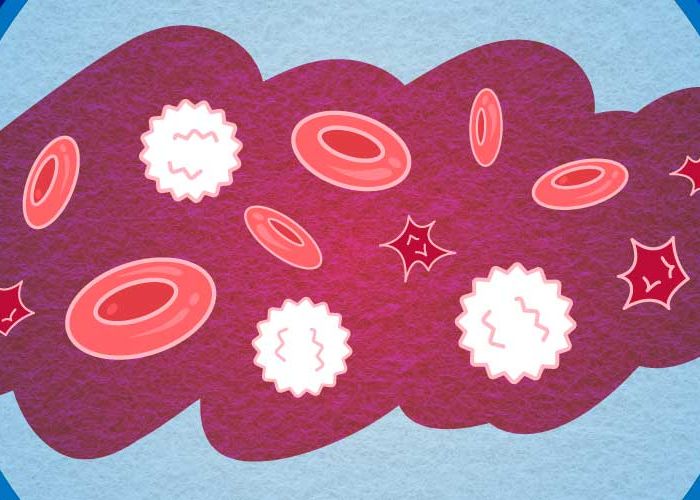
In a complete blood count (CBC), IMHA is often associated with regenerative anemia. This type of anemia is characterized by reticulocytosis and anisocytosis. Early in the disease, IMHA may appear non-regenerative because the bone marrow response might lag behind the rapid destruction of red blood cells. This highlights the importance of serial blood monitoring instead of relying on a single CBC to determine the presence of regeneration if not initially present.
If we examine the CBC more closely, two key indicators that strongly support an IMHA diagnosis are spherocytosis and autoagglutination. Spherocytes are antibody-coated red blood cells that have been partially destroyed. While a small number of spherocytes may be subtle, moderate to large numbers seen on a blood smear of an anemic dog suggest an immune-mediated disease process. Autoagglutination is the visible clumping of red blood cells caused by antibodies and is typically detected through a saline autoagglutination test.
In severe cases, patients may also show hyperbilirubinemia, hyperbilirubinuria, hemoglobinemia, hemoglobinuria, and elevated lactate levels. Although no single finding is definitive, IMHA is most often diagnosed based on a combination of clinical signs and compatible laboratory results.
Primary Versus Secondary IMHA
Another challenging aspect of this disease is distinguishing between primary (idiopathic) and secondary IMHA. Secondary IMHA occurs when the immune-mediated destruction of red blood cells is triggered by infectious diseases, inflammation, drug exposure, neoplasia, or other underlying causes. Failing to identify the underlying cause can negatively impact treatment response and prognosis.
The differential workup should include:
-
Ruling out tick-borne diseases (e.g., Ehrlichia, Anaplasma, Babesia)
-
Advanced imaging and diagnostics to rule out solid and hematopoietic tumors
-
A thorough vaccination and drug history
A diagnosis of primary IMHA should be made only after a reasonable attempt to rule out secondary causes through thorough history-taking, physical exam, and focused diagnostics.
The Role of Blood Morphology and Other Diagnostics
A key diagnostic step in the IMHA workup is a blood morphology assessment. A well-prepared blood smear allows direct visualization of spherocytes, ghost cells, and agglutination. This emphasizes the importance of not relying solely on automated hematology results but also reviewing the blood smear in every anemic patient.
Beyond blood morphology, serum biochemistry, urinalysis, and the diagnostics mentioned above, another test that may be considered is the direct antiglobulin test (Coombs' test). While a Coombs' test can be helpful in certain cases, false positives and false negatives are possible depending on the disease stage and previous immunosuppressive therapy. This test result should not be used in isolation.
As veterinarians gain more access to innovative, in-practice, point-of-care diagnostics, recognizing regenerative anemia becomes easier. Automated analyzers report reticulocyte counts and flag abnormalities, and some include a workflow with an integrated blood smear. Real-time diagnostics can greatly reduce diagnostic time and enable earlier intervention.
Early Recognition and Case Management
Unfortunately, IMHA has a guarded prognosis. Mortality rates range from 30% to 70% depending on disease severity and complications. However, evidence consistently shows that early recognition, tailored and prompt therapy, and careful monitoring are key factors in improving survival rates.
A timely and accurate diagnosis enables veterinarians to initiate immunosuppressive therapy, reduce the risk of thromboembolic complications, and provide supportive care, such as blood transfusions when needed. Because the diagnostic process for this disease can be complex and expensive, clear communication with clients is crucial. Open discussion should include the diagnostic plan to rule out secondary causes first, the tests that support immune-mediated disease, and the treatment options and costs. This transparency helps build trust, which is especially important during complex workups, long-term management, or when the prognosis is uncertain.
Final Thoughts
IMHA remains one of the most challenging diseases to diagnose and treat in small animal practice. Prompt, accurate diagnosis depends on recognizing key clinical signs and bloodwork results, systematically ruling out secondary causes, and using blood morphology along with modern diagnostic tools. By adopting standardized diagnostic workflows and emphasizing early detection, veterinarians can reduce treatment delays, improve client communication, and ultimately achieve better outcomes for patients facing this life-threatening disease.